
Imaging
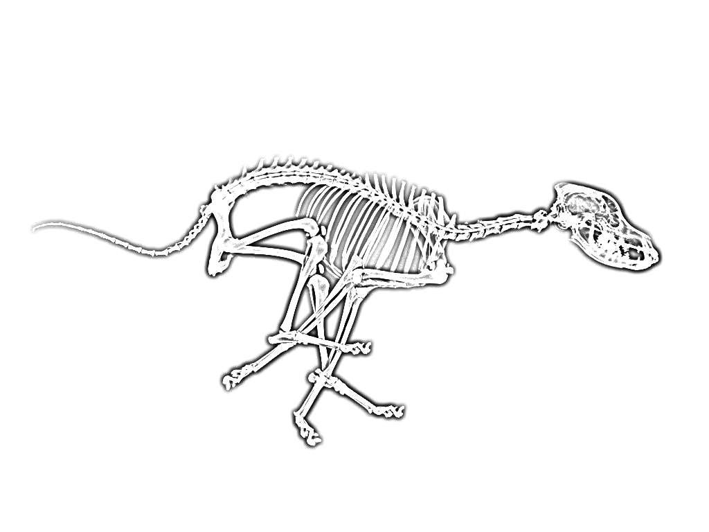
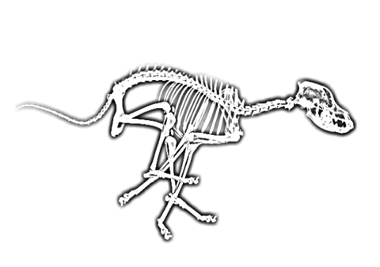
Radiography is one of the most commonly used diagnostic tools in veterinary practice. It provides a large amount of information to the veterinarian by noninvasive and economical means. It does not alter the disease process or cause unacceptable discomfort to your pet. The true advantage of digital images is that they can be easily sent via the internet to a Board Certified Veterinary Radiologist for review. Ultrasounds use sound waves and the echoes they send back to generate a digital image of the internal organs and structures in a body. Just as with humans, veterinary ultrasound can be used to scan a pet non-invasively to check for a wide variety of conditions. Ultrasound is useful for many things beyond the traditional cardiac and abdominal exams. Ultrasound can be used to evaluate muscle lesions, deep subcutaneous or intramuscular mass lesions, chronic draining tracts, the mediastinum, and even some pulmonary lesions.
At Padonia Veterinary Hospital we have one of the most sophisticated, filmless, and advanced X-Ray machine in the area. We use direct digital radiography (DR) system where the radiographic images taken from a pet are transferred to a computer and directly displayed on a digital monitor. This allows for dramatic time and effort savings over traditional film-based radiography The ultrasound at Padonia Hospital can also be used to guide a procedure used to biopsy masses and thus avoid unnecessary and more invasive surgical procedures. Because our ultrasound is also digital, it allows us to share images electronically with specialists for advanced consultation. Padonia Veterinary Hospital maintains relationships with board-certified radiologists, surgeons, and specialists in veterinary internal medicine, cardiology, oncology, and many other fields.
Our Services
-
In-Home Humane Euthanasia
-

Grooming
Health / Care -
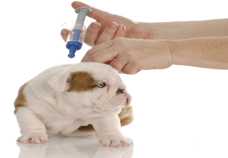
Vaccination
Health / Prevention -
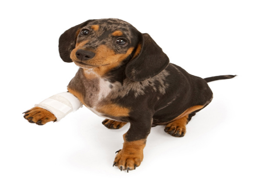
Emergency Care
Health / Care -
Flea Control
Health / Prevention / Care -
Nutritional Counseling
Fitness / Nutrition / Health -
Surgery
Health / Care -
Dental Care
Health / Prevention / Care -

Pick Up and Drop Off
Health / Prevention / Care -
Imaging
Health / Prevention / Care -

Humane Euthanasia
Health / Care -
Pain Management
Health / Prevention / Care